Calcium Imaging
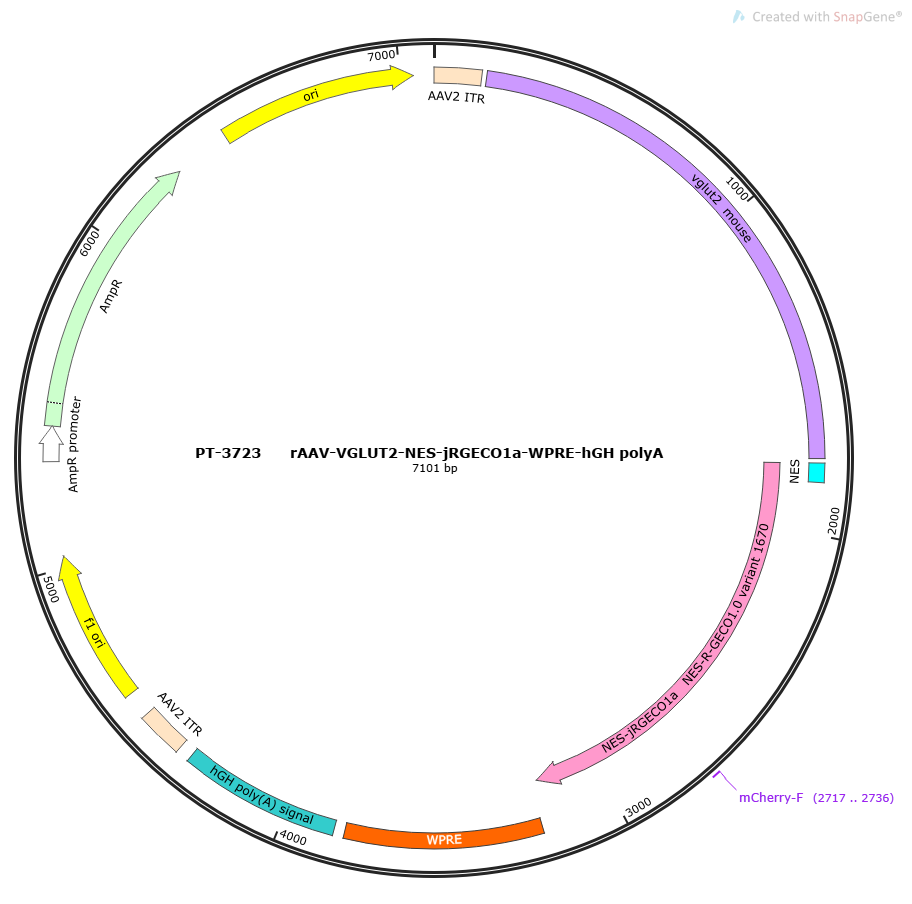
PFD-rAAV-EF1α-DIO-NES-jRGECO1a-WPRE-hGH polyA
jRGECO1a is red genetically encoded calcium indicators(GECIs). It is fusion protein composed of a calmodulin-binding peptide, a circularly permuted red fluorescent protein and calmodulin.
AAV1, AAV2, AAV3, AAV4, AAV5, AAV6, AAV8, AAV9, retro, AAV11, AAV13, PHP.eB, BR1, B10, MG1.AAV2, olig001, DJ, php.s, 9/retro
jRGECO1a
EF1α Broad-spectrum Promoter
Store at -80°C
Delivery Information
Usually Ships in 7 Business Days;
Product Details
Promoter Details
Ef1α, a strong promoter, human targeted elongation factor 1α (EF1A) gene, can be stably expressed in most types of cells, especially cells where the CMV promoter is silenced, such as stem cells, 1178bp.
Functional Gene Details
jRGECO1a: Red-shifted calcium imaging, fluorescence from mApple, NES-RCaMP1h variant 1519, active in mice, flies, fish and worms, using deep brain imaging, can be used simultaneously with the GCaMP green fluorescence series to study the activity and interactions of different types of neurons. The disadvantage is that existing red fluorescent detectors are not as useful as green ones. It can be used to study deep tissues by reducing scattering and tissue absorption, thereby reducing phototoxicity. The excitation wavelength is 1040nm or 1100nm.
jRGECO1a: Red-shifted calcium imaging, fluorescence from mApple, NES-RCaMP1h variant 1519, active in mice, flies, fish and worms, using deep brain imaging, can be used simultaneously with the GCaMP green fluorescence series to study the activity and interactions of different types of neurons. The disadvantage is that existing red fluorescent detectors are not as useful as green ones. It can be used to study deep tissues by reducing scattering and tissue absorption, thereby reducing phototoxicity. The excitation wavelength is 1040nm or 1100nm. NES: export nuclear signals, nuclear export signal sequence, only distributed outside the nucleus and located in the cytoplasm.
Fluorescent Tag
-
Fluorescent Protein Details
mCitrine: yellow fluorescent protein, excitation wavelength 516nm, emission wavelength 529nm. Tag protein: HA: The HA tag protein sequence is YPYDVPDYA, which is derived from the hemagglutinin surface epitope of influenza virus. It has 9 amino acids and has little impact on the spatial structure of the foreign target protein. It is easy to construct a tag protein fused to the N-terminus or C-terminus. Easy to detect with Anti-HA antibody detection and ELISA. IRES: internal ribosome entry site, the Chinese name is the internal ribosome entry sequence, which can be inserted downstream of the CDS region on the mRNA to mediate another CDS region that follows it, thus enabling the expression of two proteins on one mRNA.
Application
Measuring neuronal activity using genetically encoded calcium indicators (GECIs) has become a widely used method in neuroscience. Calcium imaging with GECIs is often used to measure neural activity in the complete nervous system, such as detecting the activity of a large number of neuronal cell bodies, or tracking the dynamics of neuronal subcells (such as axons, dendrites, and single synapses). Calcium imaging system is a type of optogenetic technology that uses calcium ion indicators that can sense Ca2+ concentration to detect Ca2+ concentration changes in cells or tissues and convert calcium concentration changes into fluorescent signals, thereby converting cell electrical activity into recordable light signals.
Regulatory Element Details
DIO: Double-floxed inverse Orientation, is an element that cooperates with Cre recombinase to control the expression of the target gene. If there is no Cre recombinase, the target gene will not be expressed, but in the presence of Cre, the target gene can be expressed normally. NES: export nuclear signals, nuclear export signal sequence, only distributed outside the nucleus and located in the cytoplasm.
Notes
1. Short-term storage at 4°C; long-term storage at -80°C
2. It can be stored at -80°C for more than 6 months. Each time it is frozen and thawed, the titer will be reduced by 10%, and repeated freezing and thawing should be avoided; if the virus is stored for more than 6 months, it is recommended to re-measure the virus titer before use
3. According to the experimental needs, the virus can be appropriately diluted and divided into the volume required for each experiment.
Expression and Regulation Mode
Cre-dependent