Calcium Imaging
rAAV-TRPV1-GCaMP6s-WPRE-bGH polyA
GCaMP6 is a green genetically encoded calcium indicator(GECI) tailored to detect neuronal activity, which outperformed other sensors in cultured neurons and in zebrafish, flies, and mice in vivo.High sensitive, slower kinetics, indication suitable for low frequency signals.
AAV1, AAV2, AAV3, AAV4, AAV5, AAV6, AAV8, AAV9, retro, AAV11, AAV13, PHP.eB, BR1, B10, MG1.AAV2, olig001, DJ, php.s, 9/retro
GCaMP6s
TRPV1 Neuron-specific Promoter
Store at -80°C
Delivery Information
Usually Ships in 7 Business Days;
Product Details
Promoter Details
TRPV1, mouse TRPV1 channel, pain-related promoter, 2073bp.
Functional Gene Details
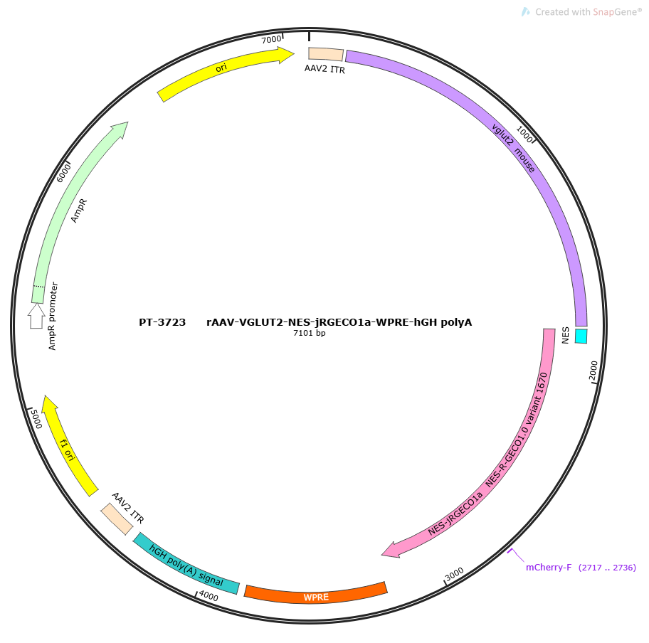
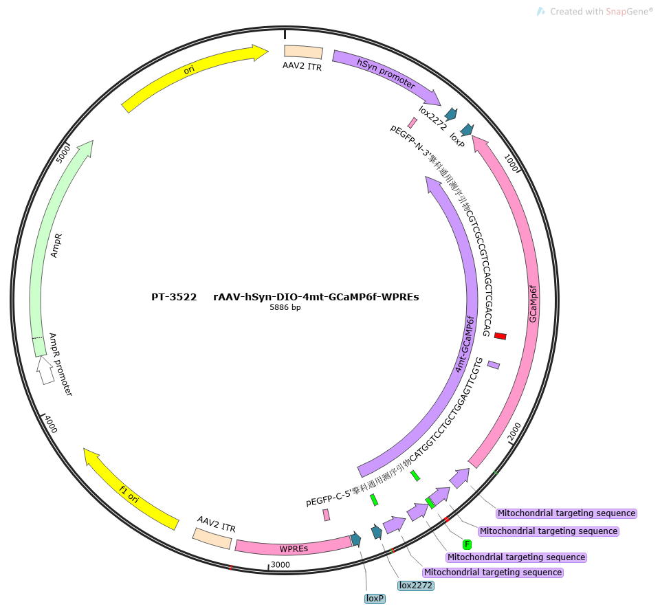
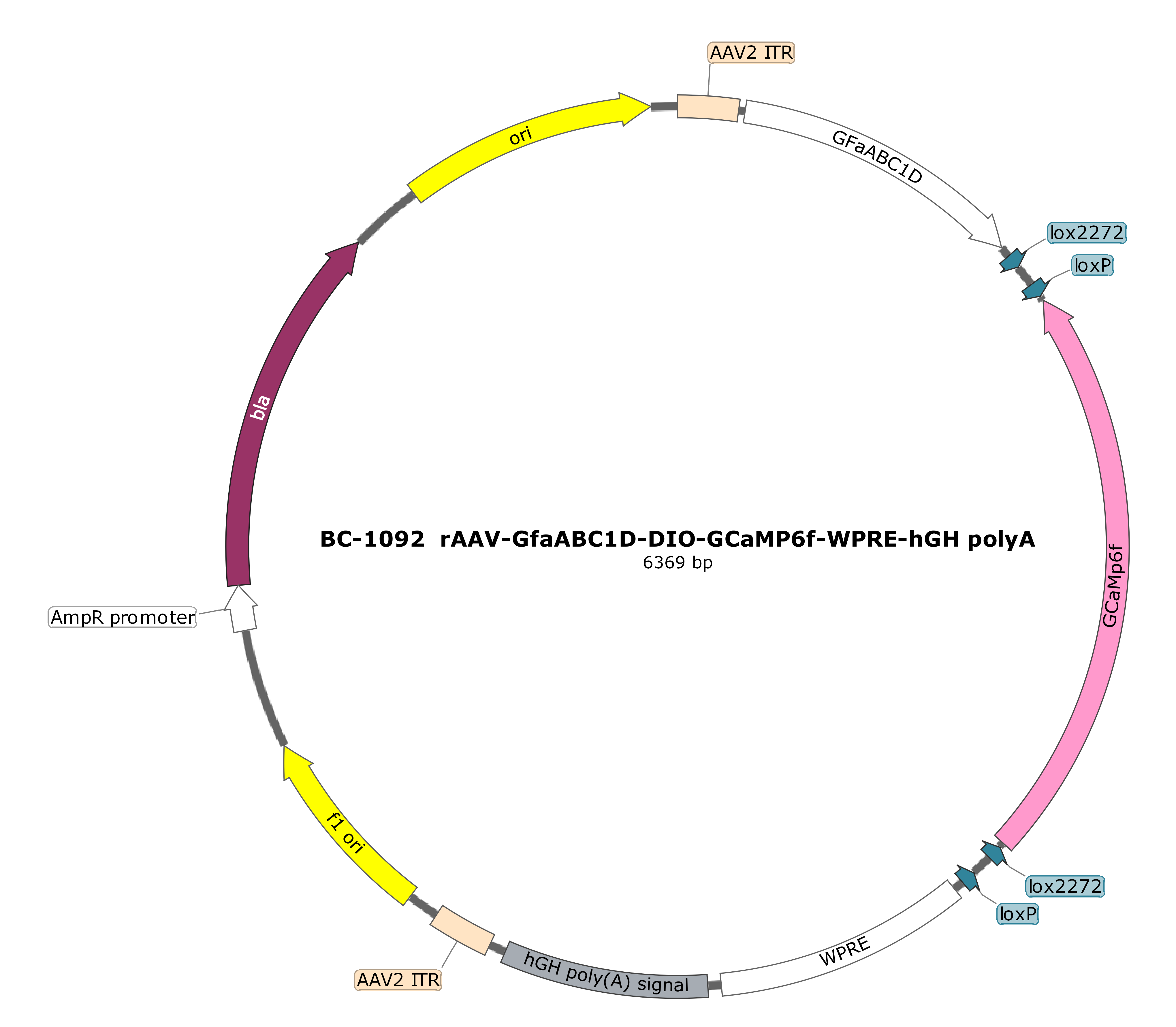
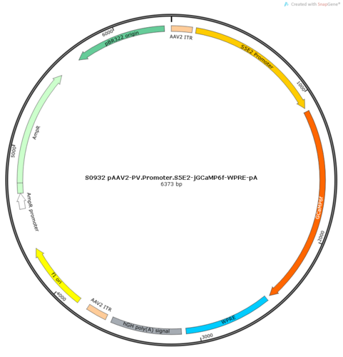
GCaMP: GCaMP consists of green fluorescent protein (GFP), calmodulin (CaM) and a peptide sequence M13 of myosin light chain kinase. When Ca2+ binds to CaM, CaM undergoes a conformational change, and its light chain region can bind to M13, causing the protonation of the chromophore of GFP to increase under illumination of a specific wavelength, and the absorbance increases, causing GFP to emit strong fluorescence. GCaMP6 is currently a conventional GECI, and its calcium indicating function reaches or even exceeds that of commonly used chemical calcium indicators. It has three commonly used subtypes, 6s, 6f and 6m. Among them, the binding force of calcium ions is 6s>6m>6f, so 6s is the most sensitive and suitable for the indication of low-frequency signals. 6f dissociates the fastest and is suitable for the indication of high-frequency signals. 6m has moderate binding activity and the widest range of application. GCaMP6 is an ultra-sensitive fluorescent protein that can specifically and efficiently reflect changes in intracellular free Ca2+ concentration. In vivo, GCaMP6 is 10 times stronger and kinetically 2 times faster than GCaMP3 for the calcium signal induced by a single action potential. Therefore, GCaMP6 fluorescent protein can be used as a hypersensitive Ca2+ probe to study the impact of external factors on Ca2+ in living cells under physiological and pathological conditions. The excitation wavelength is 495nm. GCaMP6s slow (100-150 ms) has the strongest light intensity and quenches later (most sensitive at low frequencies) GCaMP6m medium (75-100 ms) has medium light intensity and quenching (medium) GCaMP6f fast (50-75 ms) has weaker light intensity and quenches faster (high frequency)
Fluorescent Tag
——
Fluorescent Protein Details
mCitrine: yellow fluorescent protein, excitation wavelength 516nm, emission wavelength 529nm. Tag protein: HA: The HA tag protein sequence is YPYDVPDYA, which is derived from the hemagglutinin surface epitope of influenza virus. It has 9 amino acids and has little impact on the spatial structure of the foreign target protein. It is easy to construct a tag protein fused to the N-terminus or C-terminus. Easy to detect with Anti-HA antibody detection and ELISA. IRES: internal ribosome entry site, the Chinese name is the internal ribosome entry sequence, which can be inserted downstream of the CDS region on the mRNA to mediate another CDS region that follows it, thus enabling the expression of two proteins on one mRNA.
Application
Measuring neuronal activity using genetically encoded calcium indicators (GECIs) has become a widely used method in neuroscience. Calcium imaging with GECIs is often used to measure neural activity in the complete nervous system, such as detecting the activity of a large number of neuronal cell bodies, or tracking the dynamics of neuronal subcells (such as axons, dendrites, and single synapses). Calcium imaging system is a type of optogenetic technology that uses calcium ion indicators that can sense Ca2+ concentration to detect Ca2+ concentration changes in cells or tissues and convert calcium concentration changes into fluorescent signals, thereby converting cell electrical activity into recordable light signals.
Notes
1. Short-term storage at 4°C; long-term storage at -80°C
2. It can be stored at -80°C for more than 6 months. Each time it is frozen and thawed, the titer will be reduced by 10%, and repeated freezing and thawing should be avoided; if the virus is stored for more than 6 months, it is recommended to re-measure the virus titer before use
3. According to the experimental needs, the virus can be appropriately diluted and divided into the volume required for each experiment.
Expression and Regulation Mode
Others